What Is Flow Cytometry

What is flow cytometry? Flow cytometry is a semi-quantitative technique that allows you to analyze the frequency and other properties of cells stained with specific fluorochrome-conjugated antibodies. Flow cytometry is most frequently used to monitor the immune response due to the fact that the frequency and functionality of different immune cell subsets can be measured concurrently. Flow cytometers are the instrument at the center of this technique and have evolved over the past few decades to become an essential instrument for biomedical research.

To better understand flow cytometry, consider these core components of flow cytometry assays as you determine if this technique is something you should include in your research project.
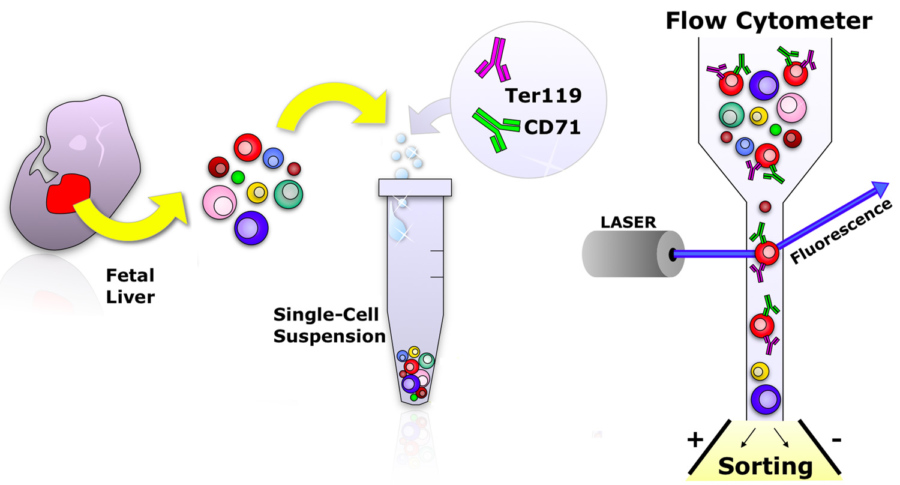
Flow cytometers can measure any single cell suspension, whether they are immune cells from the blood, tissue culture cells, or tiny beads with antibodies attached to them that can bind immune molecules like cytokines. Flow cytometry can measure multiple cell types concurrently and can discriminate between cells of different sizes and granularity.
Fluorescently labeled antibodies are the color palette for flow cytometry. Dozens of different fluorochromes or fluorochrome combinations have been engineered to attach to antibodies, be excited and emit photons at discrete wavelengths. Each antibody recognizes a specific molecule on the cell’s surface or in the cell’s interior, thus allowing detection of a specific cell population by using antibodies that bind to phenotypic markers of the cell. Each flow cytometer has unique specifications that stipulate which fluorochromes are compatible with the optics of the machine and can be detected. Now researchers regularly run experiments using flow cytometry antibody panels with 12 parameters or more, and many different fluorescent antibodies exist for immune cell markers of different species such as human, mouse, primate, etc.
Flow CytometerFlow cytometers use lasers to measure cells labeled with fluorescent antibodies. Lasers at specific wavelengths are directed at a stream of individual cells stained with fluorescent antibodies that travel through the flow cytometer in a liquid called sheath fluid. Most flow cytometers have multiple lasers that emit light at different wavelengths so a variety of fluorochromes can be measured. The laser excites fluorochrome-conjugated antibodies bound to the cells, which causes electrons from the fluorescent molecules to move to a higher energy state briefly, then return to its ground state and emit photons at a higher wavelength. Photons pass through filters so as to be measured at a specific wavelength and this signal reaches an appropriate detector, after which it is converted into voltage and reported as an event.
Computer software analyzes these events, and the data is represented to show different cell parameters including size, shape, granularity, and frequency of antibody-labeled cells. Dozens of flow cytometers exist with different lasers and emission detectors that allow use of multiple antibodies that span the visible and ultraviolet light spectra. Each cytometer has a unique configuration of excitation and emission parameters, and this must be considered when selecting antibodies for a staining panel.
